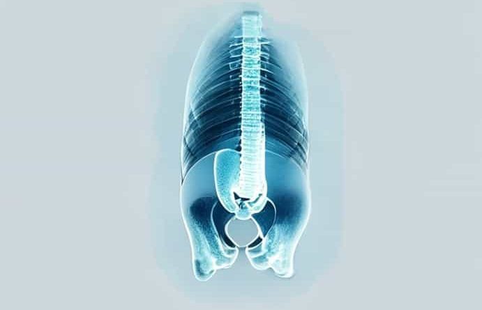
X Ray Symphysis Pubis
An X ray of the symphysis pubis is a specialized imaging procedure used to evaluate the joint located at the front of the pelvis, where the left and right pubic bones meet. This cartilaginous joint, known as the pubic symphysis, plays a critical role in maintaining pelvic stability and supporting the body’s weight during movement. X-ray imaging of this area is commonly performed to detect trauma, inflammation, degeneration, or structural abnormalities that may cause pain, mobility issues, or complications during pregnancy and childbirth. By providing detailed visualization of the pubic bones and joint space, an X ray of the symphysis pubis helps healthcare professionals make accurate diagnoses, plan treatment, and monitor recovery from injuries or chronic conditions.
Purpose of Symphysis Pubis X-ray
The primary purpose of an X ray of the symphysis pubis is to assess the integrity, alignment, and condition of the joint and surrounding bony structures. It is often requested in cases of pelvic trauma, suspected fractures, or dislocations, particularly following falls, sports injuries, or accidents. This imaging procedure is also used to evaluate conditions such as osteitis pubis, a painful inflammation of the symphysis pubis, and degenerative changes related to arthritis. Additionally, it can detect congenital abnormalities, tumors, or infections affecting the joint. In obstetric care, X-rays may be performed post-delivery if there are concerns about pelvic instability or pain, although alternative imaging methods like MRI are often preferred during pregnancy to avoid radiation exposure.
Common Indications for Symphysis Pubis X-ray
- Trauma or fractures to the pelvic region
- Chronic pelvic pain or inflammation
- Degenerative joint disease or arthritis affecting the pubic symphysis
- Osteitis pubis or sports-related pelvic stress injuries
- Detection of bone tumors or cysts in the pubic area
- Post-surgical evaluation and monitoring of pelvic reconstruction
Procedure for X-ray Symphysis Pubis
Performing an X-ray of the symphysis pubis involves precise positioning and the use of specialized imaging techniques. The patient is usually positioned supine on an X-ray table, with the legs extended or slightly rotated to optimize visualization of the joint. The technician carefully aligns the X-ray beam to focus on the symphysis pubis, capturing clear images of the joint space and surrounding bones. Lead aprons or shields may be used to protect other areas of the body from radiation exposure. Depending on the case, additional angled views may be obtained to assess subtle injuries or misalignments that might not be visible on standard frontal images. The procedure is quick, usually lasting only a few minutes, and is generally well tolerated by patients.
Types of X-ray Views
Several views may be utilized to thoroughly evaluate the symphysis pubis
- Anteroposterior (AP) ViewProvides a frontal image of the pelvis and pubic symphysis for overall alignment assessment.
- Oblique ViewsAngled perspectives to detect subtle fractures, joint irregularities, or displacement.
- Outlet and Inlet ViewsSpecialized pelvic views to examine the vertical and horizontal alignment of the pubic bones.
- Stress ViewsUsed in some cases to evaluate pelvic stability under controlled stress or movement.
Interpretation of X-ray Results
Radiologists analyze the X-ray images for abnormalities in the symphysis pubis, including widening, narrowing, fractures, or irregular bone surfaces. In cases of trauma, a fracture or diastasis may be visible as a separation or misalignment of the pubic bones. Chronic conditions such as osteitis pubis can present as joint space irregularities, sclerosis, or small bone spurs. Degenerative changes may include reduced joint space and irregular bone margins, which indicate wear and tear over time. Any abnormal findings are communicated to the referring physician, who will use the information to guide treatment, which may include rest, physical therapy, anti-inflammatory medications, or surgical intervention if necessary.
Advantages of Symphysis Pubis X-ray
- Non-invasive and relatively quick diagnostic method
- Provides clear visualization of bones and joint space
- Helps identify fractures, inflammation, or degenerative changes
- Supports treatment planning and post-injury monitoring
- Can be combined with other imaging techniques for comprehensive assessment
Safety Considerations
X-ray imaging involves exposure to a low level of ionizing radiation, which is generally considered safe when proper precautions are taken. Protective measures, such as lead shielding, help minimize exposure to surrounding tissues. It is important for patients to inform healthcare providers if they are pregnant or may be pregnant, as alternative imaging methods like MRI or ultrasound may be preferred during pregnancy. Technicians follow strict protocols to ensure minimal radiation doses while obtaining high-quality images. When used appropriately, X-ray imaging of the symphysis pubis is a safe and effective diagnostic tool for evaluating pelvic conditions.
Post-Procedure Care
After an X-ray of the symphysis pubis, there is typically no special aftercare required. Patients can resume normal activities unless advised otherwise by their healthcare provider. If the imaging was performed due to trauma or injury, follow-up care may include rest, physical therapy, or bracing to support the pelvis during recovery. Patients should report any increased pain, swelling, or unusual symptoms to their physician, as these may indicate complications or the need for additional imaging or treatment.
Conditions Diagnosed Using Symphysis Pubis X-ray
X-ray imaging of the symphysis pubis is particularly effective in diagnosing the following conditions
- Pelvic fractures and diastasis of the pubic bones
- Osteitis pubis and chronic inflammation of the joint
- Degenerative arthritis affecting the pubic symphysis
- Stress-related injuries in athletes
- Bone infections such as osteomyelitis
- Congenital or developmental pelvic abnormalities
- Post-surgical evaluation of pelvic reconstruction or hardware placement
An X-ray of the symphysis pubis is an essential diagnostic tool for evaluating pelvic stability, joint health, and bony structures in the front of the pelvis. By providing clear, non-invasive imaging, it allows healthcare professionals to detect fractures, inflammation, degenerative changes, and other abnormalities that may cause pain or mobility issues. The procedure is quick, safe, and highly informative, supporting accurate diagnosis and effective treatment planning. Whether used for trauma assessment, chronic condition monitoring, or post-surgical follow-up, X-ray imaging of the symphysis pubis remains a vital component of comprehensive pelvic care, helping patients maintain mobility, relieve pain, and ensure long-term pelvic health.