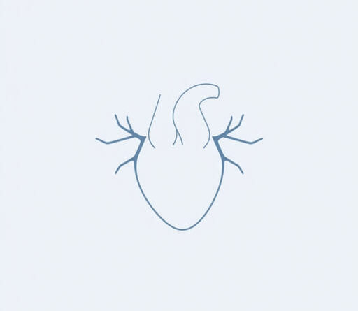
extensions of the atria
The human heart is a complex muscular organ composed of four chambers two atria and two ventricles. While the atria serve primarily as receiving chambers for blood returning to the heart, they also possess structural extensions known as auricles. These small, ear-like outpouchings are often overlooked but play an important role in cardiac anatomy and function. Understanding the extensions of the atria, particularly the auricles, is essential for students of medicine, anatomy enthusiasts, and healthcare professionals involved in cardiovascular diagnostics and treatment.
Anatomy of the Atria
Overview of Atrial Chambers
The atria are the two upper chambers of the heart the right atrium and the left atrium. The right atrium receives deoxygenated blood from the body via the superior and inferior vena cava, while the left atrium receives oxygenated blood from the lungs through the pulmonary veins. Each atrium has a muscular extension known as the auricle or atrial appendage.
Auricles: Key Extensions
Auricles are small, muscular pouches connected to each atrium. The right and left auricles are also referred to as the right atrial appendage and left atrial appendage, respectively. These structures increase the surface area of the atria and have functional and clinical importance in blood flow dynamics and rhythm regulation.
Right Atrial Extension: The Right Auricle
Structure and Position
The right auricle projects anteriorly from the right atrium and has a rough interior surface due to the presence of pectinate muscles. It lies just above the right atrioventricular (tricuspid) valve and contributes to the overall capacity of the right atrium.
Function and Relevance
The right auricle helps increase the volume of blood the right atrium can accommodate during diastole. It also contributes to atrial contraction, aiding in the movement of blood into the right ventricle. This extension plays a role in modulating venous return, particularly during increased physical activity.
- Located above the right atrioventricular groove
- Contains pectinate muscles
- Involved in atrial contraction
Left Atrial Extension: The Left Auricle
Structure and Position
The left auricle is a narrow, tubular extension of the left atrium that curves forward around the pulmonary trunk. Compared to the right auricle, it has a more slender appearance and is generally smaller in size. Like its counterpart, it also contains pectinate muscles, though these are less prominent.
Clinical Significance
The left auricle has gained significant attention in clinical cardiology due to its association with atrial fibrillation (AF). Blood can pool in the left atrial appendage during AF, leading to the formation of thrombi (blood clots). These clots can potentially dislodge and cause strokes. For this reason, the left auricle is often targeted in surgical or catheter-based procedures to prevent thromboembolic events.
- Often involved in stroke risk during atrial fibrillation
- Common site for clot formation
- May be surgically occluded or removed in certain patients
Embryological Origin of Atrial Extensions
Developmental Perspective
During embryological development, the auricles originate from the primitive atrium. As the heart matures, the smooth-walled part of the atria forms from venous inflow tracts, while the auricles retain the original muscular, trabeculated nature of the early atrium. This explains the structural and functional differences between the auricles and the main atrial chambers.
Implications for Congenital Anomalies
Developmental anomalies involving the atrial appendages are rare but can occur. Abnormalities may include accessory appendages, malformations, or structural variations that can complicate cardiac imaging and surgical planning. Awareness of embryological origins is important in interpreting these variations.
Physiological Functions of Atrial Extensions
Reservoir Function
Both auricles act as reservoirs during ventricular systole, allowing the atria to store blood temporarily. When the ventricles relax, the auricles contract, propelling blood into the ventricles and improving cardiac efficiency.
Endocrine Role
The atrial extensions are also involved in the secretion of atrial natriuretic peptide (ANP), a hormone that regulates blood volume and pressure. This hormone is released in response to atrial stretch and acts on the kidneys to increase sodium excretion, reduce blood volume, and lower blood pressure.
Electrophysiological Contributions
Auricles contain specialized muscle fibers that may contribute to the atrial conduction system. While not primary pacemakers, they can play a role in the propagation of electrical impulses across the atrial myocardium, especially in conditions that affect normal conduction patterns.
Clinical Procedures Involving Atrial Extensions
Left Atrial Appendage Occlusion (LAAO)
In patients with atrial fibrillation who are at high risk of stroke but cannot take anticoagulants, procedures such as the Watchman device implant or surgical occlusion of the left auricle may be performed. These interventions aim to prevent clot formation in the left atrial extension.
Ablation and Imaging
During catheter ablation procedures for arrhythmias, detailed imaging of the atrial extensions is crucial. CT and MRI scans help visualize auricular anatomy to guide catheters and avoid complications. Additionally, transesophageal echocardiography (TEE) is often used to assess auricular thrombus presence.
Comparative Anatomy in Other Species
Auricles Across Vertebrates
Extensions of the atria are not unique to humans. Many vertebrates, including mammals, birds, and reptiles, exhibit similar structures. While the functional roles remain consistent to enhance atrial capacity and assist in circulation there are species-specific differences in size, shape, and physiology.
Research Implications
Animal models often help in studying diseases like atrial fibrillation and developing surgical interventions involving the atrial appendages. Understanding comparative anatomy enhances our ability to translate experimental results into clinical practice.
The extensions of the atria, known as auricles or atrial appendages, are small yet vital components of the heart’s anatomy. The right auricle plays a role in expanding atrial volume and assisting in blood flow to the right ventricle, while the left auricle is critical in understanding stroke risk, especially in patients with atrial fibrillation. These extensions also serve physiological functions related to endocrine signaling, electrical conduction, and reservoir capacity. Clinically, they are relevant in diagnostic imaging, ablation procedures, and stroke prevention strategies. A comprehensive understanding of atrial extensions not only deepens our appreciation of cardiac anatomy but also supports better diagnosis and treatment of cardiovascular conditions.
