Knee Joint Ossification Centers
The development of knee joint ossification centers is an important topic for understanding normal skeletal growth, assessing childhood development, and interpreting radiographic images. Because the knee is one of the most complex joints in the human body, its ossification timeline provides valuable clues about bone maturity. By understanding how the femur, tibia, and patella form their ossification centers, clinicians, students, and curious readers can better appreciate the gradual transformation of cartilage into bone during childhood and adolescence. This explanation also helps distinguish normal variations from conditions requiring medical attention.
Overview of Knee Joint Ossification Centers
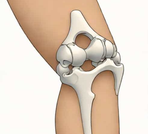
The knee joint consists of three major bones the distal femur, the proximal tibia, and the patella. Each bone develops one or more ossification centers that appear at predictable stages of childhood. These centers originate within cartilage and gradually expand, shaping the knee as a functional hinge joint. Understanding these centers is crucial for evaluating growth plates, diagnosing fractures, and assessing developmental disorders.
At birth, much of the knee is still cartilaginous. However, within the first few years of life, multiple secondary ossification centers emerge and grow until they fuse during adolescence. Although exact timing can vary between individuals, the sequence is remarkably consistent, which makes the knee useful for estimating skeletal age.
Distal Femur Ossification Center
The distal femur is typically the first part of the knee to ossify. In fact, its ossification center is often present even before birth, which makes it a useful marker in neonatal examinations. The distal femoral epiphysis expands rapidly during infancy, playing a key role in early leg growth.
Key Characteristics
- The secondary ossification center of the distal femur often appears in late fetal life or shortly after birth.
- It is the largest epiphysis in the body during growth, contributing significantly to lower limb length.
- The growth plate between the epiphysis and metaphysis remains active until adolescence.
As the distal femur grows, its ossification center gradually forms recognizable condyles. These condyles help stabilize the knee and provide smooth articulation surfaces. Because the distal femur grows so quickly, it is frequently examined by pediatric radiologists when estimating bone age or evaluating trauma.
Proximal Tibia Ossification Center
The proximal tibia ossification center usually appears very soon after the distal femur center. In many children, it emerges within the first few months of life. The tibial epiphysis forms the upper portion of the shin bone and interacts closely with the femoral condyles in the knee joint.
Key Characteristics
- The primary ossification center forms in utero, while the secondary center of the proximal tibia appears shortly after birth.
- The tibial growth plate provides a substantial portion of the tibia’s longitudinal growth during childhood.
- The tibial tuberosity, important for patellar tendon attachment, develops from a separate ossification area that appears later.
Because the tibial tuberosity has its own ossification process, it can become a site of inflammation during puberty, leading to conditions such as Osgood Schlatter disease. Understanding normal tibial ossification patterns helps prevent misdiagnosis of this region.
Patellar Ossification Centers
The patella, or kneecap, is unique because it begins entirely as cartilage. Ossification typically starts between ages three and six, although variation is common. Instead of forming from a single center, the patella often develops multiple small centers that gradually merge.
Key Characteristics
- Patellar ossification begins later than that of the femur and tibia.
- Multiple ossification centers may appear before uniting into a single bony patella.
- Incomplete fusion can result in a bipartite or multipartite patella, which is usually harmless.
Because the patella acts as a sesamoid bone within the quadriceps tendon, its ossification pattern differs from long bones. On radiographs, early patellar centers may appear fragmented or irregular, which is typically normal. Familiarity with these variations prevents confusion with fractures.
Timing and Sequence of Ossification
Although exact timing varies, the general sequence of knee joint ossification centers is highly consistent. This makes the knee an important reference point for determining skeletal age in children. Healthcare professionals often rely on radiographs to compare the development of ossification centers against standardized growth charts.
Typical Timeline
- Late fetal life or at birth Distal femur secondary ossification center appears.
- Birth to a few months Proximal tibia secondary ossification center appears.
- Around age 3 to 6 Patella begins to ossify from multiple small centers.
- Early adolescence Tibial tuberosity ossification becomes more pronounced.
- Mid to late adolescence Growth plates of the femur and tibia begin to close.
By the late teenage years, the knee ossification centers are normally fully fused, marking the end of growth. The sequence and development of these centers are used in forensic studies, pediatric orthopedics, and sports medicine.
Importance in Clinical Evaluation
Understanding knee joint ossification centers is essential for interpreting pediatric radiographs. When evaluating injuries such as fractures, clinicians must distinguish between growth plates, accessory ossification centers, and true bone disruptions. Misinterpreting normal developmental features can lead to unnecessary concern or incorrect treatment.
Knowledge of ossification patterns also plays a vital role in diagnosing developmental conditions. Delayed ossification may signal endocrine disorders, nutritional deficiencies, or genetic conditions. Conversely, unusually early ossification may indicate hormonal imbalances. Regular monitoring helps ensure that children’s growth follows a typical trajectory.
Common Variations
While the general pattern of knee ossification is consistent, some variations are frequent and usually harmless. These variations can appear unusual to the untrained eye but are well-documented in medical literature.
Notable Variations
- Bipartite patellaOccurs when the patella has two ossification centers that do not fuse completely; typically asymptomatic.
- Accessory ossification centersSmall, additional centers may appear around the knee and eventually fuse or remain separate without causing problems.
- Tibial tuberosity fragmentationCommon during adolescence and often part of normal development.
Recognizing these variations helps avoid confusion with fractures or pathology. When symptoms do appear, they are usually related to irritation rather than structural defects.
Knee joint ossification centers provide a fascinating look into the body’s growth process. From the early appearance of the distal femur center to the gradual formation of the patella, each stage contributes to the development of a stable and functional joint. Understanding these centers is valuable for clinicians, students, and anyone interested in human anatomy. The predictable sequence of ossification assists with age estimation, injury interpretation, and evaluation of developmental disorders. By appreciating the complexity and timing of these changes, we gain deeper insight into how the knee grows, adapts, and supports the body through childhood and adolescence.