Knee Joint Ossification Centres
The development of the knee is a fascinating and highly organized process, and one of the most important aspects of that development involves the appearance and growth of knee joint ossification centres. These centres form the foundation for the future structure of the distal femur, proximal tibia, and patella. Because the knee joint is essential for weight-bearing, mobility, and stability, understanding how its early bone formation occurs can help explain later differences in growth, posture, and even certain medical conditions. Parents, students, and health enthusiasts often explore this topic to better understand how bones transition from soft cartilage into the strong tissue that supports walking, running, and daily activity.
What Ossification Centres Are
Ossification centres are specific points where bone tissue begins to form inside cartilage or membrane. In the knee joint, these centres play a major role in shaping the final anatomy. Humans are born with many bones still partially made of cartilage, which allows flexibility during birth and provides room for rapid growth after delivery. As the body matures, ossification continues in a predictable pattern.
In general, there are two types of ossification centres
-
Primary ossification centres, which appear during prenatal development
-
Secondary ossification centres, which typically appear after birth
The knee joint contains both types, and their timing can be important for assessing bone age, identifying growth delays, or understanding how injuries affect children differently from adults.
Main Knee Joint Ossification Centres
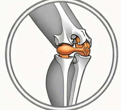
The knee involves three major regions the distal femur, the proximal tibia, and the patella. Each develops its own ossification centre or centres. The timeline varies slightly between individuals, but the general pattern remains consistent enough to be used in pediatric assessment.
Distal Femur Ossification Centre
The distal femur is known for having one of the earliest and most prominent secondary ossification centres in the body. In many cases, this centre is already visible at birth. Its presence is often used by pediatric radiologists to help determine neonatal bone maturity.
The distal femur ossification centre lies just above the knee joint. As it expands, it shapes the future femoral condyles, which play a crucial role in smooth joint movement. The early appearance of this centre is important because the femur must begin supporting weight soon after a child’s first steps.
Proximal Tibia Ossification Centre
The proximal tibia also forms a major secondary ossification centre shortly after birth. This centre influences the growth of the tibial plateau, a surface critical for joint stability and weight distribution.
Over time, the growth plate between the tibial shaft and the secondary ossification centre remains active through childhood and adolescence. This growth plate is one of the reasons children are more vulnerable to specific injuries, such as tibial tuberosity avulsion. As ossification progresses, the final shape of the knee joint becomes more refined, supporting improved coordination and mobility.
Patellar Ossification Centres
The patella, or kneecap, is unique because it develops from several ossification centres that eventually fuse. Unlike the femur and tibia, the patella begins as a cartilaginous structure entirely separate from the other bones of the knee. Ossification usually starts between ages three and six.
These centres grow toward each other until they merge into a single solid bone. Variants can occur, such as bipartite or multipartite patella, where the centres fail to fuse completely. Although usually harmless, these variations can occasionally cause knee pain, especially in active individuals.
Why Timing of Ossification Matters
The timing of knee joint ossification centres offers valuable information about a child’s development. Healthcare professionals often use radiographic imaging to evaluate bone age, which provides insight into growth rate and maturity. If a centre appears earlier or later than expected, it may indicate a difference in development.
Several factors influence ossification timing
-
Genetics
-
Nutrition, especially calcium and vitamin D intake
-
Hormonal levels
-
Chronic illness or metabolic disorders
While minor variations are normal, significant delays or accelerations may prompt further evaluation. In sports medicine and orthopedics, understanding the activity of these centres helps guide decisions about training intensity, injury risk, and treatment options tailored to growing bones.
Growth Plates and Their Relationship to Ossification
Growth plates, or epiphyseal plates, are regions of cartilage where new bone is produced. They remain active until late adolescence. In the knee, growth plates are located near the ends of the femur and tibia, close to the secondary ossification centres.
The interplay between ossification centres and growth plates determines how long bones lengthen. Damage to a growth plate can disrupt bone development, sometimes resulting in asymmetry or altered joint alignment. Because the knee contains some of the largest growth plates in the body, childhood knee injuries require careful assessment.
Common Conditions Related to Knee Ossification Centres
Although the appearance of ossification centres is a natural part of growth, certain conditions can affect them. Awareness of these issues helps parents and students understand why regular monitoring may be necessary.
Osgood-Schlatter Disease
This condition affects the area where the patellar tendon attaches to the tibia. When the growth plate near the tibial tuberosity becomes irritated, it can cause swelling and pain. Rapid growth combined with athletic activity increases the risk.
Bipartite Patella
When patellar ossification centres fail to fuse completely, a bipartite patella can form. Most people feel no discomfort, but some may experience symptoms during jumping, running, or kneeling.
Growth Plate Injuries
Accidents or sports impacts can injure the growth plates near the knee. Because these areas are still developing, the same force that causes a simple sprain in an adult may cause a more complex injury in a child.
How Healthcare Professionals Use Ossification Information
Doctors, radiologists, and physical therapists rely on knowledge of knee ossification centres to diagnose conditions, track development, and create effective treatment plans. For example, the presence or absence of certain centres in X-rays can help determine a child’s skeletal age, which may differ from their chronological age. This information guides decisions about growth expectations, potential interventions, and activity limitations.
Orthopedic specialists also monitor ossification when evaluating leg alignment. Knock knees, bow legs, and other alignment patterns can be normal phases of growth, but if ossification patterns appear unusual, they can signal an underlying issue.
How Ossification Shapes Movement and Strength
The gradual hardening of bone in the knee joint affects how a child moves. As ossification centres enlarge and growth plates thicken, the knee becomes more capable of absorbing force and maintaining stability. This process supports transitions from crawling to walking, then running, jumping, and more complex movements.
Because each centre contributes to a specific part of the knee’s anatomy, balanced development is essential. For example, the distal femur shapes the smooth rolling surface of the knee joint, while the patella improves leverage for the quadriceps muscles. All of these elements depend on proper ossification.
The development of knee joint ossification centres is a remarkable example of how the human body grows from flexible cartilage into strong, functional bone. The predictable appearance of these centres in the distal femur, proximal tibia, and patella allows professionals to assess growth, identify disorders, and guide treatment for children and adolescents. Understanding these centres also helps explain why growing knees behave differently from adult joints, especially in sports, injury response, and overall mobility. With proper awareness and monitoring, most children experience smooth and healthy development as their knees mature and their bones reach adult strength.