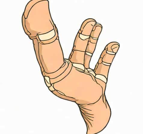
Ulnar Base Of Proximal Phalanx
The ulnar base of the proximal phalanx is a critical anatomical structure within the human hand, playing a significant role in finger movement, stability, and overall hand function. Situated at the base of the proximal phalanx on the ulnar, or medial, side of the finger, this area serves as a key attachment point for ligaments and tendons that facilitate flexion, extension, and intricate motions. Understanding the ulnar base of the proximal phalanx is essential for healthcare professionals, including orthopedic surgeons, physiotherapists, and hand therapists, as injuries or deformities in this region can significantly impair hand function. This topic explores the anatomy, biomechanics, clinical relevance, and rehabilitation considerations associated with the ulnar base of the proximal phalanx, providing a comprehensive overview for both professionals and students of anatomy.
Anatomical Overview
The proximal phalanx is the first bone in each finger following the metacarpal, connecting the metacarpophalangeal (MCP) joint to the middle phalanx. The ulnar base refers specifically to the portion of the proximal phalanx that is located on the ulnar side, which corresponds to the side of the hand aligned with the little finger. This region is characterized by several important anatomical landmarks, including small ridges and depressions that serve as attachment points for collateral ligaments and tendinous insertions. The surface anatomy is subtle but crucial for proper biomechanical function, as it ensures stability during lateral stresses and contributes to the fine motor movements necessary for grasping and manipulating objects.
Ligamentous Attachments
One of the most important aspects of the ulnar base of the proximal phalanx is its ligamentous connections. The ulnar collateral ligament (UCL) attaches to this region, providing medial stability to the MCP joint. The integrity of the UCL is essential for resisting valgus forces, which occur when the finger is pushed outward away from the midline of the hand. Damage to the UCL at the ulnar base of the proximal phalanx can lead to instability, decreased grip strength, and deformities such as a skier’s thumb in the case of the thumb, or similar injuries in the fingers. Understanding the precise anatomy of these ligament attachments is critical for surgical repair and rehabilitation planning.
Tendinous Insertions
In addition to ligaments, the ulnar base of the proximal phalanx also serves as an insertion point for tendons of intrinsic and extrinsic muscles of the hand. The flexor and extensor tendons pass near this region, and some fibers of the interosseous muscles insert on the ulnar side, contributing to fine motor control and finger abduction/adduction. These tendinous attachments allow for coordinated movement and dexterity, which are essential for daily activities such as writing, typing, or holding utensils. Injury to these tendinous insertions can result in limited range of motion and functional deficits.
Biomechanical Importance
The ulnar base of the proximal phalanx plays a vital role in the biomechanics of the finger. During flexion and extension, this region functions as a fulcrum for ligamentous and tendinous structures, transmitting forces efficiently between the phalanges and metacarpals. The ulnar base also helps stabilize the finger against lateral forces, preventing deviation that could compromise grip and pinch strength. Its contribution to the complex kinematics of the finger is especially apparent during precision tasks that require controlled lateral movement or when the hand is subjected to external stresses, such as in sports or manual labor.
Load Distribution
Another key function of the ulnar base is distributing load across the MCP joint. When the finger grips or manipulates objects, forces are transmitted through the phalanges to the metacarpals. The ulnar base of the proximal phalanx helps absorb and redirect these forces, reducing stress on the joint capsule and articular cartilage. This load distribution is essential for preventing long-term degenerative changes, such as osteoarthritis, and maintaining optimal hand function over time.
Clinical Relevance
Injuries to the ulnar base of the proximal phalanx can occur from trauma, overuse, or degenerative processes. Common injuries include ligament tears, avulsion fractures, and tendon injuries. Avulsion fractures, in particular, involve the detachment of a small fragment of bone where ligaments or tendons attach, often caused by sudden forceful movements. Recognition of these injuries is critical, as failure to properly treat them can result in chronic instability, loss of function, and pain.
Diagnosis and Imaging
Diagnosis of ulnar base injuries typically involves clinical examination and imaging studies. Physical tests may include stress tests for collateral ligament integrity, palpation for tenderness, and assessment of range of motion. Imaging modalities such as X-rays, MRI, or ultrasound provide detailed visualization of bone, ligament, and tendon involvement. Accurate diagnosis allows healthcare providers to determine the best course of treatment, which may range from conservative management with splinting and therapy to surgical intervention for severe or complex injuries.
Surgical and Non-Surgical Management
Non-surgical treatment of minor injuries at the ulnar base of the proximal phalanx often involves immobilization, anti-inflammatory medication, and hand therapy. Splinting ensures stability while soft tissues heal, and targeted exercises help restore strength and range of motion. Surgical repair may be required for complete ligament tears, significant avulsion fractures, or chronic instability. Procedures typically aim to reattach ligaments or tendons to the ulnar base, restore alignment, and ensure functional recovery. Post-operative rehabilitation is critical, focusing on gradual mobilization, strengthening, and coordination exercises to regain full hand functionality.
Rehabilitation Considerations
Effective rehabilitation after an injury to the ulnar base of the proximal phalanx emphasizes both passive and active mobilization. Hand therapists often design programs that target the recovery of flexion, extension, and lateral stability. Specific exercises may include controlled range of motion drills, grip strengthening, and fine motor skill training. Attention to proprioception is also important, as the ulnar base contributes to the hand’s ability to sense position and movement. Consistent therapy helps prevent stiffness, maintain joint health, and restore the intricate functions necessary for daily tasks.
Preventive Measures
Preventing injuries to the ulnar base of the proximal phalanx involves awareness of proper hand positioning and ergonomic practices. Athletes and workers who perform repetitive hand motions may benefit from protective taping, strength training, and regular stretching to reduce the risk of ligament or tendon damage. Education on safe techniques for gripping, lifting, and manipulating objects can further minimize the likelihood of trauma to this sensitive region of the hand.
The ulnar base of the proximal phalanx is a small yet highly significant component of hand anatomy, integral to stability, movement, and overall hand function. Its ligamentous and tendinous attachments, biomechanical contributions, and role in load distribution make it essential for both everyday activities and specialized tasks. Injuries in this area can lead to substantial functional impairment, emphasizing the importance of accurate diagnosis, appropriate management, and targeted rehabilitation. Understanding the anatomy and clinical significance of the ulnar base of the proximal phalanx is crucial for healthcare professionals, hand therapists, and anatomy students alike. By recognizing its role in stability, dexterity, and force transmission, we can better appreciate the intricate design of the human hand and the delicate balance that allows it to perform complex, coordinated movements.
Ultimately, the ulnar base of the proximal phalanx exemplifies how even small anatomical structures have profound implications for function and health. Attention to this area in both clinical practice and hand therapy ensures optimal recovery, preserves hand dexterity, and supports the intricate coordination required for everyday life.