Label The Parts Of The Ruminant Stomach
The ruminant stomach is a fascinating and complex organ that allows animals like cows, sheep, goats, and deer to digest fibrous plant material efficiently. Unlike monogastric animals that have a single-chambered stomach, ruminants possess a multi-chambered stomach, which enables them to extract nutrients from cellulose-rich foods. Labeling and understanding the parts of the ruminant stomach is essential for students of veterinary science, animal husbandry, and biology. Each chamber has a specific function, contributing to the breakdown of food, fermentation, absorption, and nutrient assimilation. By examining the structure and function of these stomach compartments, we can gain a deeper appreciation for the ruminant digestive system.
Overview of the Ruminant Stomach
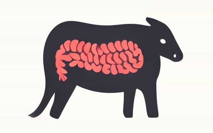
The ruminant stomach consists of four distinct compartments the rumen, reticulum, omasum, and abomasum. These compartments work together to process large quantities of fibrous plant material that would be indigestible to many other animals. The ruminant digestive system also involves chewing cud, a process called rumination, which allows the animal to regurgitate partially digested food and chew it again to facilitate further breakdown. Each stomach compartment has a specialized structure and function, and labeling these parts is essential to understanding how ruminants efficiently utilize roughage.
Rumen
The rumen is the largest compartment of the ruminant stomach and serves as the primary site for microbial fermentation. It occupies the left side of the abdominal cavity and can hold large volumes of ingested food. The inner lining of the rumen contains papillae, which increase the surface area for nutrient absorption. Microorganisms such as bacteria, protozoa, and fungi live in the rumen and break down complex carbohydrates like cellulose into volatile fatty acids, which provide energy to the animal. Labeling the rumen on a diagram typically highlights its position at the front and left of the stomach complex.
Reticulum
The reticulum is the second chamber and is located adjacent to the rumen, often described as having a honeycomb structure. Its primary function is to trap heavy or dense objects, such as stones or metal, preventing them from causing injury to the digestive tract. The reticulum also participates in the fermentation process alongside the rumen and plays a critical role in initiating regurgitation during rumination. Labeling the reticulum emphasizes its close connection to the rumen and its distinct honeycomb lining, which distinguishes it from the other stomach compartments.
Omasum
The omasum is the third compartment of the ruminant stomach, sometimes referred to as the manyplies due to its multiple folds. These folds increase the surface area for absorption of water, minerals, and volatile fatty acids. The omasum acts as a filter, reducing the ptopic size of ingested food before it enters the abomasum. Its muscular walls also help grind the food and regulate its passage. When labeling the omasum, attention is given to its spherical shape and the folded structure, which are characteristic features important for its function.
Abomasum
The abomasum is the fourth and final compartment of the ruminant stomach and is often called the true stomach. Unlike the previous compartments, the abomasum functions similarly to the monogastric stomach, secreting digestive enzymes and acids that break down proteins and other nutrients. It prepares the digesta for further digestion and absorption in the small intestine. Labeling the abomasum highlights its position as the terminal compartment of the ruminant stomach and its role in enzymatic digestion.
Importance of Labeling the Ruminant Stomach
Accurately labeling the parts of the ruminant stomach is crucial for understanding animal nutrition, health, and veterinary care. Each compartment plays a unique role in digestion, and knowledge of their functions helps in diagnosing digestive disorders, formulating proper diets, and improving livestock management practices. Students and professionals often use diagrams to visualize the location, structure, and interconnections of the rumen, reticulum, omasum, and abomasum.
Applications in Animal Husbandry
Understanding the ruminant stomach is important for optimizing feeding strategies. For example, the rumen requires a balanced population of microorganisms to ferment fibrous feed effectively. Supplements, probiotics, and feed management practices are designed to maintain rumen health. Recognizing the roles of the reticulum, omasum, and abomasum helps in addressing problems such as hardware disease, impactions, or poor nutrient absorption. Labeling diagrams of the stomach can serve as an educational tool for farmers and veterinary students to identify potential issues and implement corrective measures.
Educational Tools and Techniques
Labeling exercises for the ruminant stomach often include diagrams, 3D models, and even dissections. These tools help students visualize the size, shape, and position of each compartment. Key points to focus on when labeling include
- The rumen’s large volume and papillae-covered lining
- The reticulum’s honeycomb structure and proximity to the rumen
- The omasum’s folded walls and role in absorption
- The abomasum’s enzymatic activity and position as the true stomach
Such exercises reinforce understanding of the ruminant digestive system and provide practical knowledge applicable to both academic and professional settings.
Common Misconceptions
Many people mistakenly believe that ruminants have a single stomach or that all compartments function identically. Labeling the stomach accurately clarifies these misconceptions by showing the distinct structure and specialized function of each part. Another common error is confusing the reticulum with the rumen, since they are closely connected and often function together in fermentation and cud formation. Clear labeling helps distinguish these compartments and emphasizes their individual contributions to digestion.
Visual Learning Benefits
Using labeled diagrams enhances retention and understanding of complex anatomical structures. By associating each compartment with its specific function, students can better grasp how the ruminant stomach efficiently processes fibrous plant material. Visual learning is particularly effective for remembering the sequence of compartments and their interrelated roles in digestion, rumination, and nutrient absorption.
The ruminant stomach is a complex, multi-chambered organ designed for efficient digestion of fibrous plant material. Labeling its four parts-the rumen, reticulum, omasum, and abomasum-helps students, farmers, and veterinary professionals understand the structure and function of each compartment. The rumen serves as a fermentation chamber, the reticulum traps foreign objects and aids in regurgitation, the omasum absorbs water and nutrients, and the abomasum carries out enzymatic digestion. Understanding these roles is essential for animal nutrition, health management, and educational purposes. By studying and labeling the ruminant stomach, we gain insight into the remarkable adaptations that allow these animals to thrive on a plant-based diet, emphasizing the interplay between anatomy, physiology, and agricultural practices.