Treatment Of Epidural Hematoma
Epidural hematoma is a serious medical condition that occurs when blood accumulates between the dura mater, the outer protective layer of the brain, and the skull. This type of hematoma is typically caused by head trauma, often resulting from accidents, falls, sports injuries, or violent impacts. Epidural hematomas can lead to increased intracranial pressure, compression of brain tissue, and, if left untreated, life-threatening complications. Prompt recognition and timely treatment are crucial to prevent permanent brain damage and improve outcomes. Understanding the causes, symptoms, diagnostic methods, and available treatment options is essential for managing epidural hematomas effectively and ensuring patient safety.
Understanding Epidural Hematoma
An epidural hematoma occurs when blood vessels, usually arteries, are torn due to trauma, leading to rapid accumulation of blood in the epidural space. This condition often develops quickly and can cause a range of neurological symptoms, including headaches, confusion, dizziness, nausea, vomiting, and loss of consciousness. Because the bleeding occurs between the skull and dura mater, pressure builds on the brain tissue beneath, which can compromise brain function. Early intervention is essential to reduce the risk of long-term complications or death.
Common Causes
Epidural hematomas are most commonly associated with head injuries that result in skull fractures or direct trauma to the temporal region. Key causes include
- Motor vehicle accidents, which are a leading cause of traumatic brain injury.
- Falls, especially in elderly individuals or those with impaired mobility.
- Sports injuries, including contact sports such as football, hockey, or boxing.
- Physical assaults or violent impacts to the head.
- Skull fractures that damage the middle meningeal artery, which is often involved in arterial epidural hematomas.
Symptoms and Warning Signs
The symptoms of an epidural hematoma can vary depending on the size and location of the hematoma and the speed of blood accumulation. Classic signs include a lucid interval, where the patient initially appears conscious and alert after injury, followed by deterioration in mental status. Other warning signs include
- Severe headache that worsens over time.
- Nausea and vomiting.
- Confusion, agitation, or drowsiness.
- Weakness or numbness on one side of the body.
- Seizures or convulsions.
- Pupil changes, such as dilation or unequal pupils, indicating increased intracranial pressure.
Diagnosis of Epidural Hematoma
Timely and accurate diagnosis is critical for effective treatment of epidural hematomas. Medical professionals use a combination of physical examination, patient history, and imaging studies to confirm the condition.
Neurological Assessment
Healthcare providers perform a detailed neurological examination to evaluate consciousness, pupil response, motor function, and sensory perception. This helps determine the severity of brain compression and guides treatment decisions.
Imaging Studies
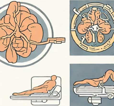
Imaging is essential for diagnosis. Computed tomography (CT) scans are the preferred method due to their ability to quickly identify the location, size, and extent of bleeding. Magnetic resonance imaging (MRI) may be used in specific cases to provide more detailed images of brain tissue and surrounding structures.
Treatment Options for Epidural Hematoma
The treatment of an epidural hematoma depends on the size of the hematoma, the severity of symptoms, and the patient’s overall condition. Rapid medical intervention is often required to prevent permanent neurological damage.
Surgical Treatment
Surgery is the primary treatment for most epidural hematomas, especially when the hematoma is large or causing significant neurological symptoms. Surgical options include
- Craniectomy or CraniotomyA portion of the skull is removed to access and remove the accumulated blood, relieving pressure on the brain.
- Burr Hole EvacuationSmall holes are drilled into the skull to drain the hematoma, typically used for smaller or less severe hematomas.
Surgery is often performed as an emergency procedure, as rapid decompression can prevent further brain injury and improve patient outcomes.
Non-Surgical Management
In select cases where the hematoma is small and the patient is stable, careful monitoring may be sufficient. Non-surgical management includes
- Frequent neurological assessments to detect changes in consciousness or motor function.
- Use of medications to control intracranial pressure, such as osmotic diuretics (e.g., mannitol) or hypertonic saline.
- Pain management and supportive care, including oxygen therapy and bed rest.
- Follow-up imaging to ensure the hematoma does not enlarge or cause complications.
Post-Treatment Rehabilitation
Recovery from an epidural hematoma may require rehabilitation, depending on the severity of the injury and the patient’s neurological status. Rehabilitation focuses on restoring cognitive, physical, and functional abilities.
Physical Therapy
Physical therapy helps patients regain strength, balance, coordination, and mobility. Exercises are tailored to the individual’s needs and may include motor skill training, balance exercises, and endurance building.
Occupational Therapy
Occupational therapy assists patients in relearning daily activities and improving fine motor skills. Therapy focuses on restoring independence in tasks such as dressing, eating, and personal care.
Cognitive and Speech Therapy
If the hematoma or increased intracranial pressure caused cognitive or speech deficits, therapy may include memory exercises, problem-solving strategies, and speech-language rehabilitation to regain communication skills.
Prevention and Safety Measures
While not all epidural hematomas can be prevented, certain safety measures reduce the risk of head trauma and subsequent hematoma formation.
Use of Protective Gear
Wearing helmets during sports, cycling, and motor vehicle activities can significantly reduce the risk of head injuries that lead to epidural hematomas.
Fall Prevention
For elderly individuals, implementing home safety measures such as handrails, non-slip mats, and proper lighting can prevent falls and head trauma.
Safe Driving Practices
Using seat belts, following traffic rules, and avoiding distracted or impaired driving help prevent motor vehicle accidents that often result in epidural hematomas.
When to Seek Emergency Medical Attention
An epidural hematoma is a medical emergency. Immediate care should be sought if any of the following symptoms occur after head trauma
- Severe headache that worsens rapidly.
- Loss of consciousness or confusion.
- Nausea, vomiting, or seizures.
- Weakness or numbness on one side of the body.
- Changes in pupil size or vision disturbances.
- Any sudden deterioration in alertness or coordination.
Treatment of epidural hematoma requires prompt recognition, accurate diagnosis, and timely intervention. Surgical evacuation through craniotomy or burr holes is often necessary for large hematomas or significant neurological compromise, while smaller hematomas may be managed with careful monitoring and supportive care. Post-treatment rehabilitation, including physical, occupational, and cognitive therapy, is essential for restoring function and quality of life. Preventive measures such as helmet use, fall prevention, and safe driving practices reduce the risk of head trauma and subsequent epidural hematomas. Recognizing early symptoms and seeking immediate medical attention are critical steps to minimize complications and improve outcomes. Through comprehensive treatment and rehabilitation strategies, individuals affected by epidural hematomas can achieve optimal recovery and regain independence in daily activities.